Age Related Macula Degeneration
AMD or Age-related Macula Degeneration is currently the leading cause of sight loss in the western world and is becoming more of a problem as medical advances enable us to living longer. There are different stages to AMD and two different types of the disease, both are explained in more detail below. Dry AMD is a very slow progressive condition whereas Wet AMD is much more aggressive and affects the vision greatly very quickly. Currently there are more treatment options available for Wet AMD but there is advice available to try and prevent the disease from affecting you. Before we get to the detail of AMD lets have a look at the normal retinal structure and how the retina works. This will help you to understand what exactly is happening in the AMD eye.
Just click any image on this page to enlarge
The Retina
The retina is a marvel of evolutionary bio-engineering. By it's very nature it is a highly sensitive structure which is susceptible to attack from harmful substances and even light (the wrong kind that is).
|
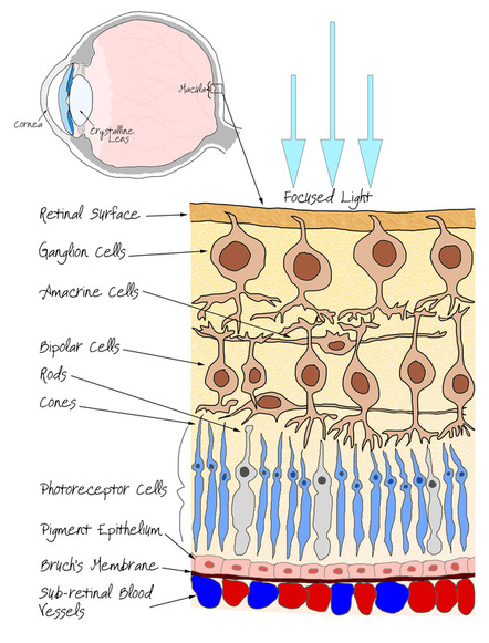
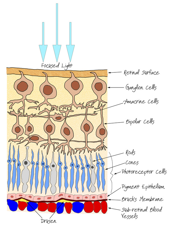
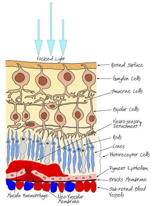
The normal retinal structure can be seen adjacent. Think of it as the film in a camera, except the circuitry of the camera is sitting on top of the film. The light enters the eye and is focused by the cornea and the crystalline lens onto the retinal surface where it passes through the retinal nerve fibres and activates both the rod and cone cells (named after their shape), resulting in a electrical signal being conducted through the nerve cells (the ganglion, bipolar and amacrine cells) on it's way to the Occipital cortex within the brain enabling us to see the world around us. As you can see the retina is very much part of the nervous system and is a peripheral component of the brain. Just like in a brain injury, should the worst happen and the retinal tissues be damaged in any way then the tissue does not repair itself and simply leaves a scar behind. In the retina, scarred tissue is blind. Think of the damage as clumps of pixels not working on your computer screen. The screen will produce an image but that image will have patchy areas of no signal.
|
You can experience retinal sensitivity for yourself by switching the lights off in a dark room. Instantaneously the room will go pitch black and your vision will take a few minutes to be restored. This is because there are only certain light sensitive cells (the rods) within the retina which can see in low light levels. In the higher illumination they are completely bleached out and over-stimulated, this means that when they are needed for low-light viewing it takes them roughly about 30 minutes to recover from the over-stimulation (or light damage) from when the room lights were on. Conversely, you may notice that once you are adapted to the low light levels, if you look directly at a small object it will disappear. This is because the light sensitive cone cells are predominantly found in the central vision and not so many rod cells. Because the cones are not sensitive enough to see in low light levels it can cause a small blind spot in the centre of your vision in lower light levels. The cone cells also provide the colour vision that we see; you may also notice that in a dark room you cannot decipher the colour of objects, again this is because the cone cells are not providing you with the information as they are not sensitive enough to see in low lighting conditions.
Thankfully the body has developed protective mechanisms and structures to help prevent any undue damage to the retina rods and cones but what if these processes degenerate with age or disease?
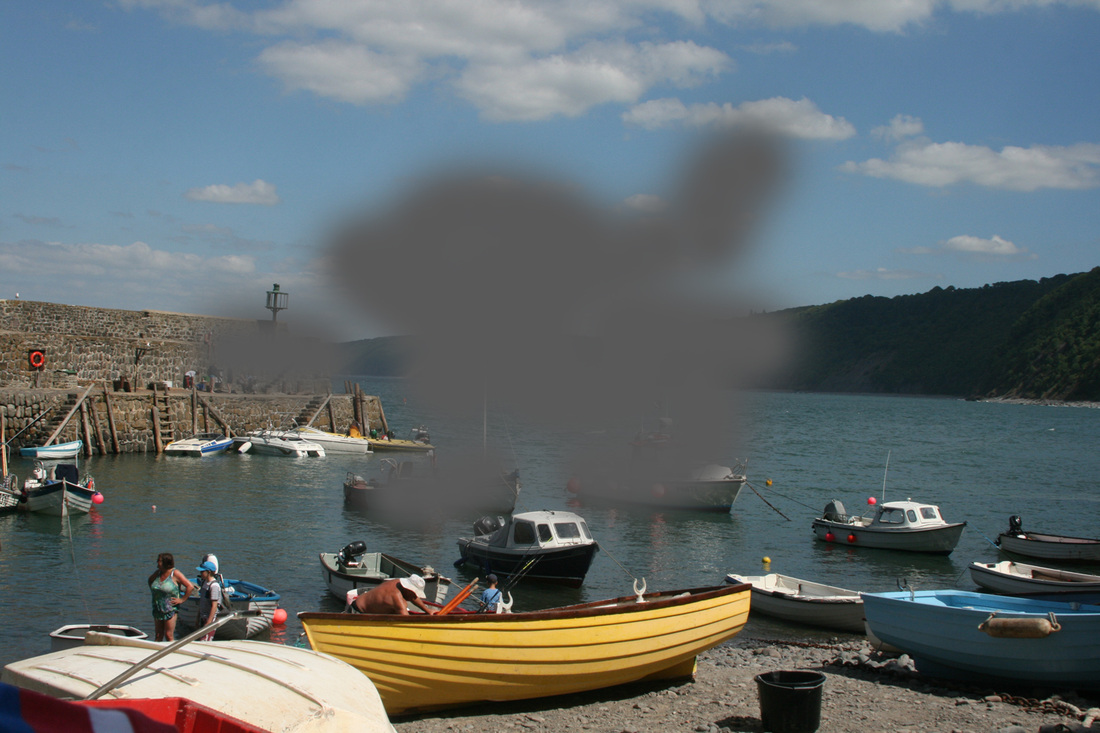
Because the macula is the area of the retina that is most highly sensitive with lots of rod and cone cells packed together, it is used for the detailed central vision, the part we use when inspecting something, reading, looking at peoples faces or watching television. When the AMD damage occurs it does not affect the peripheral retina, therefore patients with AMD will NEVER go completely blind from the condition, they will always be able to see out of the corner of the eye. What AMD does affect is the central portion of the vision, when a patient with AMD looks directly at something it will disappear, rather like the dark room experiment suggested above.
What causes AMD?
Unfortunately the answer to this question is "we don't really know fully". What we do know, however, is the risk factors. We do know that UV light, smoking and poor nutrition are all significant risk factors. Racial and gender specific risks are also evident. By far the largest group exposed to AMD is caucasian females, we believe this mainly to be a result of UV light exposure, caucasians have a smaller proportion of melanin pigment in their tissues. Melanin is instrumental in mitigating the harmful effects of UV light. Blood pressure, cholesterol and obesity have all been linked to an increased risk of AMD. Those who have a family member with AMD have also been shown to have a greater risk of developing the disease.
The current thinking is that a specific type of particle call a free radical attacks the retinal tissue progressively over you lifetime eventually causing irreversible damage resulting in AMD. Free radicals are formed by oxidants which can be found in all sorts of food and drink such as tea, coffee, particularly in tobacco smoke. They can also be formed by the activation of UV light. Cumulative exposure to these free radicals eventually causes damage to the retinal cells. For more information on ways to try and prevent AMD please scroll to the bottom section of this page.
The current thinking is that a specific type of particle call a free radical attacks the retinal tissue progressively over you lifetime eventually causing irreversible damage resulting in AMD. Free radicals are formed by oxidants which can be found in all sorts of food and drink such as tea, coffee, particularly in tobacco smoke. They can also be formed by the activation of UV light. Cumulative exposure to these free radicals eventually causes damage to the retinal cells. For more information on ways to try and prevent AMD please scroll to the bottom section of this page.
Dry AMD
|
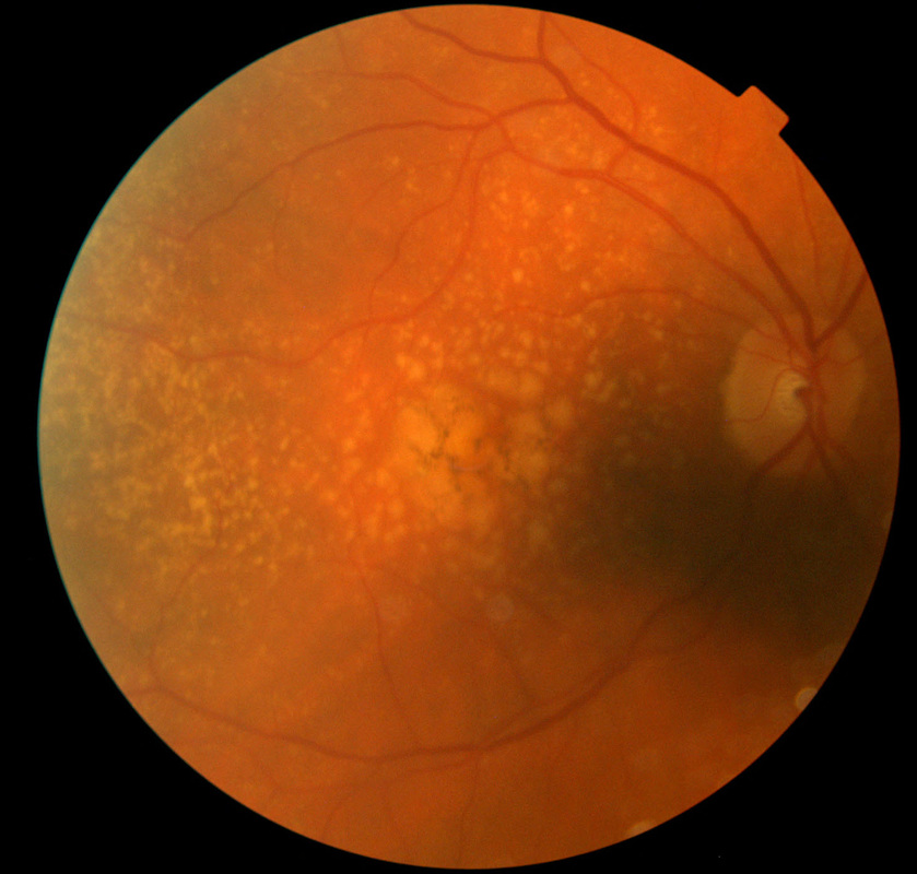
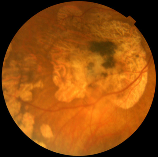
Dry AMD is a very slow and progressive condition and accounts for almost 90% of those with AMD. It is characterised by the slow atrophy or death of the photoreceptor cells (the rods and cones).
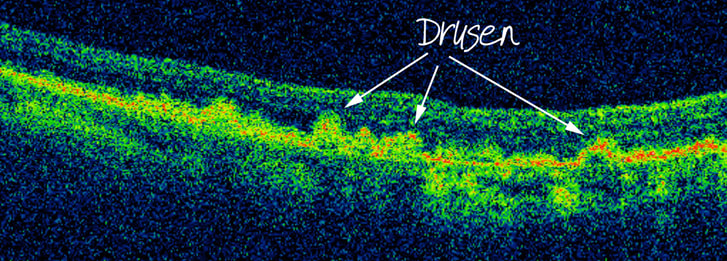
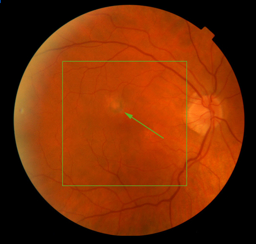
In Dry AMD the photoreceptor support structure called Bruch's Membrane thins or breaks down resulting in damage and death to the photoreceptors. The earliest sign of Dry AMD is small fluffy yellow/white spots in the retina seen at the macula called Drusen, they consist of Lipid, a fatty like substance. It is thought that they are created when the retina fails to adequately dispose of the waste products formed by the chemical reactions in the photoreceptor cells by the process of capturing the light information for your sight. Over time the waste product builds up and eventually harms the photoreceptor cells causing their death. The presence of Drusen does not automatically mean that you vision is affected and they can be present without visual disturbance for many years. They may start out as small distinct and scattered spots but as they enlarge and coalesce then they pose a greater risk of sight loss and progression to the Wet form of the disease. As the Drusen enlarge they can disrupt the layers of the retina resulting in a pigment epithelial detachment as outlined below. As for treatment, unfortunately there is currently no treatment for Dry AMD. There is advice regarding prevention of progression (please see below) and lots of organisations can offer help and support for those in need. Many people cope very well with their vision. It is important to remember that AMD is NOT a blinding condition and with a little change in routine many can cope very well. Magnifiers, although cumbersome can offer a vital lifeline and modern technology has provided a plethora of gadgets and innovations to help those affected. Advice and demonstrations can be sought via one of our Optometrists or the local Blind Society, N-Vision at Squires Gate (click the image below for more information). |
Pigment Epithelial Detachment (PED)
|
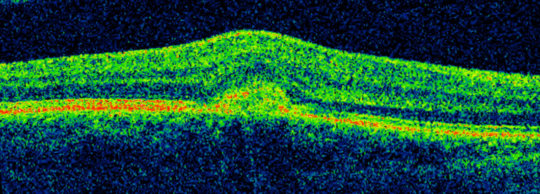
Pigment Epithelial detachment or PED occurs when there is a separation between the support structure called Bruch's membrane and the pigment epithelial layers of the retina. They can occur for a variety of reasons, not all associated with AMD. Clinically they are seen as small domed elevations within the retina usually caused by the presence of large coalesced drusen or by the growth of new blood vessels through Bruch's membrane into the inter-cellular space between the two layers.
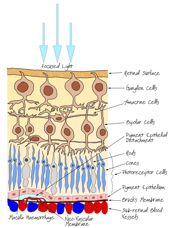
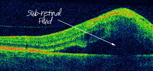
If caused by a neo-vascular membrane (new vessel growth) then there may be sub-retinal fluid associated with the PED. New blood vessels are particularly subject to haemorrhages, they are very leaky pipes, which can result in a pocket of fluid forming within the retina. This is a very early stage of Wet AMD, named after the fluid association. The raised area of tissue then disrupts the retinal structure and affects the ability of the photorecepters to function properly. Treatment of a PED is not always necessary and they can be highly resistant therapy. The PED itself is not treated but the underlying cause can be. Any fluid build up can be targeted with an appropriate anti-VegF agent. These are a group of drugs that target new-blood vessels and hinder their formation, resulting in an absorption of the intra-retinal fluid. Some practitioners advocate the use of laser treatment for PED's, this again is treating the blood vessel formation but in a much more destructive way. The laser is used to burn the surrounding retina destroying it, since a dead retina does not need oxygen or nutrients the new blood vessel growth is no longer needed resulting in resolution of the PED. This should only used when the laser treatment itself would not threaten the central vision. Upon resolution the retinal structure can be mostly restored to normal helping the photoreceptors to function better improving the vision to much better levels. |
Wet AMD
|
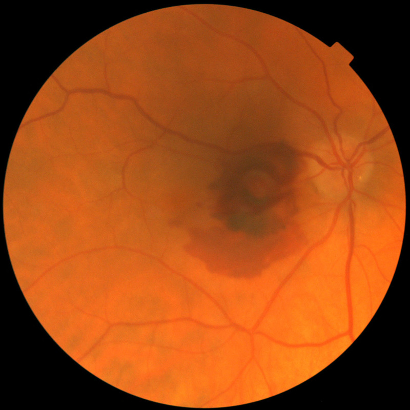
Wet AMD is named as such due to the presence of fluid within the retina. This is a sudden event arising from haemorrhaging new blood vessels (neo-vascular membrane) which has grown through Bruch's Membrane. The sudden nature of this event results in a sudden loss of vision centrally and can be quite significant.
The haemorrhage causes a neuro-sensory detachment where the photoreceptors detach from the nutrient giving support structures of the Pigment Epithelium and Bruch's Membrane and can be damaged beyond repair. The most significant symptom preceding a Wet AMD event is that the central vision becomes distorted, sometimes significantly. Objects known to be straight such as chair legs or door frames appear to be "kinked" (See the test yourself page). This is because the photoreceptor layer has become distorted by the haemorrhaging (see opposite). In recent years there has been major advancements in the treatment of Wet AMD. Anti-VegF treatments such as Lucentis (Ranibizumab) have offered real hope to those suffering with the disease. Anti-VegF agents inhibit the growth of new blood vessels and promote absorption of any intra-retinal fluid. In a large proportion of patients vision is restored to manageable levels. It is very important that the eye is treated as soon as possible once a Wet AMD event has occurred. The photoreceptors will not survive for long separated from the retinal support structures for long, therefore, the quicker the disease is diagnosed and treated then the more photoreceptors can be saved and the greater possibility of a good visual outcome. We currently operate a FAST-TRAK macula referral scheme where we notify the Hospital Eye Service directly who then have a protocol to assess any relevant patients within two weeks of referral. An older treatment known as PDT or Photo-Dynamic Therapy uses a laser to destroy the new blood vessels causing the bleed. This is a destructive treatment, however, meaning that the retina will also be destroyed around the area of bleed. It is only used where the visual outcome is not expected to improve and aims to slow or stop any progression of the disease. If you feel that you may have distorted central vision please test yourself with the Amsler grid (click here) and contact the practice ASAP or seek further medical advice. |
OCT Macula Scanning
|
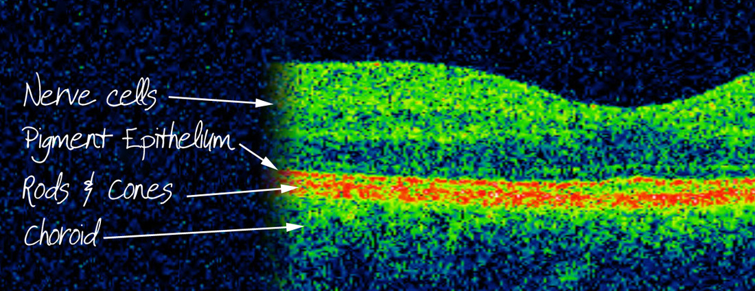
The role of OCT scanning technology is invaluable in both the early detection, diagnosis and management of AMD. It offers a level of detail that is unsurpassed by any other and is used as the gold standard in determining whether Anti-VegF treatment is indicated. The process of scanning the macula is quick and painless and anything from early stage AMD to the extent of sub-retinal fluid in a Wet AMD event can be clearly seen within minutes.
|
We highly recommend that those who have risk factors associated with AMD or are concerned about developing the condition to have a screening OCT scan at their routine eye examination as this will enable us determine whether any further advice or investigation is necessary.
Please see the OCT section for more information.
Please see the OCT section for more information.
Preventative and Nutritional Advice
|
Prevention is better than cure! The best advice to prevent, as much as possible, the disease from affecting you is as follows:
It is important to mention at this point that is is known that an increased intake of Beta-carotene increases the prevalence of lung cancer in smokers. The most important message about risk with AMD is to cease smoking and in particular, do NOT take nutritional supplements for AMD if you are a smoker. |
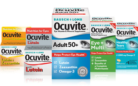
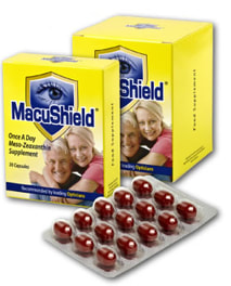
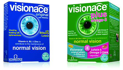
Green leafy vegetables, in particular spinach & kale, are a very good natural source of of Leutin. If you are more inclined to look for a specifically prepared formulation of the recommended nutrients there are a number of brands available some of which are outlined below. (click to images for links to information websites)
The science behind these formulations is, in the main, theoretical. However, there is one large study group based in the USA which has attempted to develop a more scientific approach. The AREDS and subsequently AREDS2 studies have been running for some time now. They have shown that those taking the AREDS2 formulation have a 20-30% lowered risk of developing AMD. This may not seem a large percentage, however, the studies are limited in both subject group size and time scale meaning that the results may be more of conservative estimate and the actual risk reduction may be much higher. We take the view, however, that any reduction in the risk should not be taken lightly as the treatment options, especially for Dry AMD, are currently very limited. We would certainly recommend that Caucasian females over the age of 50 should consider taking these supplements and also those for those with early stage disease or those who have a family member with the condition.
The science behind these formulations is, in the main, theoretical. However, there is one large study group based in the USA which has attempted to develop a more scientific approach. The AREDS and subsequently AREDS2 studies have been running for some time now. They have shown that those taking the AREDS2 formulation have a 20-30% lowered risk of developing AMD. This may not seem a large percentage, however, the studies are limited in both subject group size and time scale meaning that the results may be more of conservative estimate and the actual risk reduction may be much higher. We take the view, however, that any reduction in the risk should not be taken lightly as the treatment options, especially for Dry AMD, are currently very limited. We would certainly recommend that Caucasian females over the age of 50 should consider taking these supplements and also those for those with early stage disease or those who have a family member with the condition.
If you are at all concerned please contact the practice so that one of our Optometrists can advise you of the best course of action. See the section on Emergency Eyecare for more information on how we can help.

